The Lower Part Of Leg: Foundation for Stability and Dynamic Movement
Often taken for granted until pain or injury strikes, the lower part of leg is a marvel of biomechanical engineering. This crucial segment of our anatomy, situated elegantly between the knee and the ankle, is far more than just a connection point. It's a complex interplay of bones, muscles, tendons, nerves, and blood vessels that collectively bear our body weight, facilitate dynamic movement, and play an indispensable role in our overall mobility and stability. Understanding its intricate design and function is not only fascinating but also vital for maintaining health, preventing injuries, and appreciating the incredible capabilities of the human body.
From the powerful propulsion needed for sprinting to the subtle adjustments required for maintaining balance on uneven terrain, the structures within the lower part of leg work in concert. This article delves into the anatomical architecture of this region, explores its critical functions in movement and stability, and highlights its significant clinical importance in diagnosing and treating a wide array of conditions.
Anatomy of the Lower Part Of Leg: A Structural Masterpiece
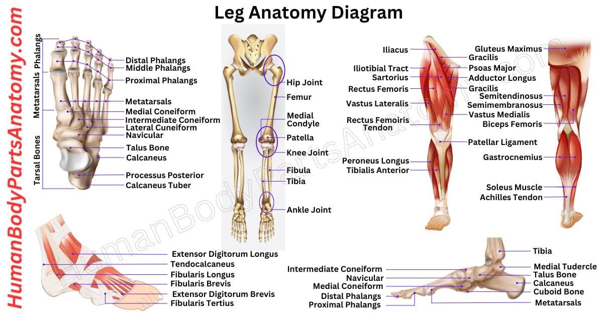
The core framework of the lower part of leg consists of two primary bones: the robust tibia and the slender fibula. These bones, along with their surrounding soft tissues, are organized into distinct compartments, each with specialized roles. This intricate arrangement allows for both immense strength and remarkable flexibility.
Bones and Joints: The Skeletal Foundation
- The Tibia (Shin Bone): As the larger and more medial of the two lower leg bones, the tibia is the primary weight-bearing bone. Its sturdy shaft extends from the knee to the ankle, forming the prominent "shin" at the front of the leg. The tibia is crucial for transmitting forces from the thigh to the foot and is a common site for fractures, especially in high-impact sports.
- The Fibula: Positioned laterally to the tibia, the fibula is significantly thinner and plays a lesser role in weight-bearing. However, its importance cannot be understated. It serves as a vital attachment point for numerous muscles of the lower leg and forms the lateral malleolus, a key component of the ankle joint that provides crucial stability. While often overshadowed by the tibia, the fibula's contribution to muscle mechanics and ankle integrity is profound.
Together, the distal ends of the tibia and fibula articulate with the talus bone of the foot to form the ankle joint. This hinge joint, reinforced by strong ligaments, is incredibly sophisticated, allowing for dorsiflexion (lifting the foot) and plantarflexion (pointing the toes), as well as more subtle inversion and eversion movements.
Muscles and Compartments: Powering Movement and Support
The muscular anatomy of the lower part of leg is organized into four main compartments, each housing specific muscle groups, nerves, and blood vessels. This compartmentalization is essential for efficient function and also has significant clinical implications:
- Anterior Compartment: Located at the front of the leg, this compartment contains muscles responsible for dorsiflexion of the foot (lifting the foot upwards) and extension of the toes. The most prominent muscle here is the tibialis anterior, crucial for walking and preventing "foot drop."
- Lateral Compartment: Situated on the outer side of the leg, this compartment houses muscles like the fibularis (peroneus) longus and brevis. These muscles primarily evert the foot (turning the sole outwards) and assist in plantarflexion, contributing significantly to balance and stability on uneven surfaces.
- Superficial Posterior Compartment (The Calf): This is the bulky, visible part of the back of the lower leg, commonly known as the calf. It contains the powerful gastrocnemius and soleus muscles, which merge to form the Achilles tendon, the strongest tendon in the body. These muscles are primary movers for plantarflexion (pushing off the ground during walking, running, and jumping).
- Deep Posterior Compartment: Lying beneath the superficial calf muscles, this compartment contains muscles that primarily invert the foot (turning the sole inwards) and assist in plantarflexion and toe flexion. Key muscles here include the tibialis posterior, vital for arch support, and the long flexors of the toes.
Beyond muscles, an intricate network of arteries, veins, and nerves ensures the vitality and function of the lower leg. The major arteries (e.g., anterior and posterior tibial arteries) supply oxygen-rich blood, while veins (e.g., great and small saphenous veins) return deoxygenated blood. Nerves (e.g., common fibular nerve, tibial nerve) transmit signals for movement and sensation, making this region incredibly responsive to its environment.
The Lower Part Of Leg's Role in Stability and Dynamic Movement
The coordinated action of the bones, joints, and muscles in the lower part of leg is fundamental to virtually every aspect of our mobility. It's a critical nexus for:
- Weight Bearing: The tibia's robust structure efficiently transfers the body's weight from the femur to the talus, enabling us to stand upright and endure significant loads during activities like running or jumping.
- Balance and Proprioception: Muscles like the tibialis anterior and the fibularis group, along with sensory receptors in the joints and tendons, provide constant feedback to the brain about the leg's position in space. This proprioceptive awareness is essential for maintaining balance, especially on unstable surfaces or during rapid changes in direction. Without the fine-tuned control offered by the lower leg, simple tasks like standing still would become incredibly challenging.
- Locomotion: The powerful calf muscles are the primary engines for propulsion, allowing us to push off the ground during walking, running, and jumping. The anterior compartment muscles then lift the foot, preventing us from tripping. This continuous cycle of muscle contraction and relaxation drives gait, making smooth and efficient movement possible.
- Shock Absorption: During impact activities, the muscles and connective tissues of the lower leg, particularly those around the ankle, act as natural shock absorbers, dissipating forces and protecting the knee and hip joints from excessive stress.
From a biomechanical perspective, the lower leg acts as a critical lever system. The muscles generate force, which is then transmitted through the tendons to move the bones at the ankle and foot joints, creating the powerful and precise movements essential for human locomotion.
Clinical Importance and Common Conditions Affecting the Lower Part Of Leg
Given its complex structure and constant use, the lower part of leg is susceptible to a variety of injuries and conditions, making its anatomical understanding crucial for healthcare professionals. Knowledge of lower limb anatomy is vital for assessing gait, diagnosing musculoskeletal injury, and interpreting orthopaedic and vascular conditions.
Musculoskeletal Injuries:
- Shin Splints (Medial Tibial Stress Syndrome): A common overuse injury characterized by pain along the inner edge of the tibia. Often seen in runners or individuals starting new exercise routines, it's typically caused by repetitive stress on the muscles and connective tissues surrounding the tibia. Proper footwear, gradual training increases, and strengthening the lower leg muscles are key to prevention and recovery.
- Stress Fractures: Small cracks in the bone, most commonly the tibia, resulting from repetitive force and overuse. They require rest and careful management to heal properly.
- Calf Strains/Tears: Injuries to the gastrocnemius or soleus muscles, often occurring during sudden acceleration or changes in direction. These can range from mild pulls to severe tears, causing significant pain and limited mobility.
- Achilles Tendonitis/Rupture: Inflammation or tearing of the Achilles tendon, the large tendon connecting the calf muscles to the heel bone. A rupture is a debilitating injury requiring immediate medical attention.
- Compartment Syndrome: A serious condition where increased pressure within one of the leg's muscle compartments compromises blood flow and nerve function. It can be acute (often due to trauma) or chronic (exercise-induced).
Vascular and Neurological Concerns:
- Deep Vein Thrombosis (DVT): Blood clots forming in the deep veins of the lower leg. This is a potentially life-threatening condition as clots can travel to the lungs (pulmonary embolism). Swelling, pain, and redness in the calf are warning signs.
- Peripheral Artery Disease (PAD): Narrowing of the arteries in the lower limbs, reducing blood flow. This can cause pain during walking (claudication) and, in severe cases, lead to ulcers and tissue death.
- Nerve Entrapments: Nerves in the lower leg can become compressed, leading to pain, numbness, tingling, or weakness. Sciatica, for example, can manifest with symptoms radiating into the lower leg.
For clinicians, understanding the precise location of nerves, blood vessels, and muscle origins/insertions in the lower part of leg is paramount for accurate diagnosis, effective surgical planning, and targeted rehabilitation strategies. Imaging interpretation (X-rays, MRI, ultrasound) relies heavily on a solid grasp of this regional anatomy.
Maintaining Optimal Lower Leg Health
Given the pivotal role the lower part of leg plays in our daily lives, proactive care is essential. Here are some actionable tips:
- Strength Training: Incorporate exercises that strengthen both the calf muscles (calf raises) and the shin muscles (tibialis anterior raises) to maintain muscular balance.
- Flexibility: Regular stretching of the calf muscles, Achilles tendon, and hamstrings can improve range of motion and reduce injury risk.
- Proper Footwear: Wear shoes that provide adequate support and cushioning, especially during physical activity. Replace worn-out athletic shoes regularly.
- Gradual Progression: When starting new exercise programs or increasing intensity, do so gradually to allow your lower leg tissues to adapt and avoid overuse injuries.
- Listen to Your Body: Pay attention to persistent pain or discomfort. Rest, ice, compression, and elevation (RICE) can help with minor issues, but seek medical advice for severe or lingering symptoms.
- Hydration and Nutrition: Support overall tissue health with a balanced diet and sufficient water intake.
Conclusion
The lower part of leg is an unsung hero of human locomotion and stability. Its complex interplay of bones, muscles, and neurovascular structures allows us to stand, walk, run, and perform countless daily activities with grace and power. From providing a stable base to propelling us forward, its functions are indispensable. A comprehensive understanding of its anatomy and biomechanics is not only fascinating but also critical for preventing injuries, addressing common conditions, and promoting lifelong mobility. By appreciating its intricate design and adopting proactive health practices, we can ensure this vital segment of our body continues to support our active lives for years to come.