The lower part of the leg is a marvel of biomechanical engineering, a complex interplay of bones, muscles, nerves, and blood vessels that enables us to stand, walk, run, and jump. Far more than just a connecting segment, this vital region—situated between the knee and the ankle—bears significant body weight, provides stability, and is crucial for locomotion. Understanding its intricate anatomy is not only fascinating but also fundamental for athletes, healthcare professionals, and anyone interested in maintaining mobility and preventing injury.
This comprehensive guide delves into the foundational structures of the lower part of the leg, exploring its skeletal framework, the powerful muscle groups that drive movement, and the key anatomical regions that define it. We'll also touch upon its clinical significance, offering insights into why this area is so important for overall strength, balance, and athletic performance.
The Bony Framework of the Lower Part Of Leg
At the core of the lower part of the leg are two distinct bones, working in conjunction to provide structure, support, and attachment points for muscles. These bones are the tibia and the fibula.
The Tibia: The Weight-Bearer
Often referred to as the shin bone, the tibia is the larger and more medial (inner) of the two lower leg bones. It's the primary weight-bearing bone in the lower part of the leg, transmitting forces from the femur (thigh bone) down to the foot. Its robust structure is essential for supporting the entire body's weight during standing and movement.
- Proximal End: Articulates with the femur to form the knee joint, featuring two prominent condyles (medial and lateral) for this articulation.
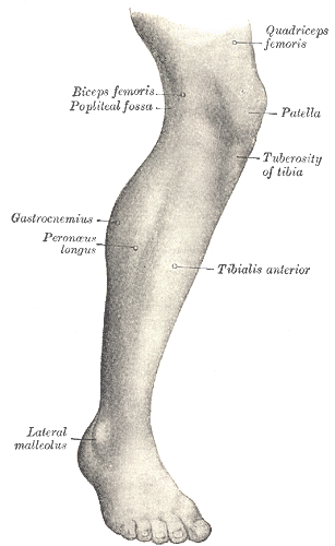
- Shaft: A triangular prism shape, with a sharp anterior border that is easily palpable, forming the "shin" of the lower part of the leg.
- Distal End: Expands to form the medial malleolus, a bony prominence on the inner side of the ankle, which forms part of the ankle joint and helps stabilize it.
The Fibula: Stability and Muscle Attachment
The fibula is the thinner, more lateral (outer) bone of the lower part of the leg. Unlike the tibia, it plays a relatively minor role in weight-bearing. Its primary functions are to provide crucial attachment sites for numerous muscles of the lower part of the leg and to contribute to the stability of the ankle joint.
- Proximal End: Articulates with the tibia just below the knee, but does not directly articulate with the femur.
- Shaft: A slender bone with various ridges and surfaces for muscle attachments.
- Distal End: Forms the lateral malleolus, the prominent bony knob on the outer side of the ankle. This structure extends further distally than the medial malleolus, providing critical lateral stability to the ankle joint and protecting it from excessive inversion.
The Interosseous Membrane
Connecting the tibia and fibula along their lengths is a strong sheet of fibrous tissue known as the interosseous membrane. This membrane plays a vital role in binding the two bones together, distributing forces, and providing additional surface area for muscle attachments. It's a critical component in maintaining the structural integrity of the lower part of the leg.
Muscular Powerhouses: Compartments of the Lower Leg
The muscles of the lower part of the leg are organized into distinct fascial compartments, each containing specific muscle groups, nerves, and blood vessels. This compartmentalization is key to their specialized functions and is also clinically important, as issues like compartment syndrome can arise when pressure builds up within these confined spaces.
The Anterior Compartment: Dorsiflexion and Toe Extension
Located at the front of the lower part of the leg, this compartment is primarily responsible for dorsiflexion of the foot (lifting the foot upwards towards the shin) and extending the toes. Key muscles include:
- Tibialis Anterior: The largest muscle in this compartment, crucial for dorsiflexion and inversion of the foot. Its strength is vital for walking and preventing foot drop.
- Extensor Digitorum Longus: Extends the four lateral toes and assists with dorsiflexion.
- Extensor Hallucis Longus: Extends the great toe (hallux) and also assists with dorsiflexion.
- Fibularis Tertius: A small muscle, often considered part of the Extensor Digitorum Longus, that assists in dorsiflexion and eversion.
Problems in this compartment, particularly overuse of the tibialis anterior, are often implicated in conditions like shin splints (medial tibial stress syndrome), a common issue for runners and athletes.
The Lateral Compartment: Eversion Specialists
Situated on the outer side of the lower part of the leg, this compartment houses muscles primarily responsible for eversion of the foot (turning the sole outwards). They also contribute to plantarflexion.
- Fibularis Longus (Peroneus Longus): Runs down the outer leg, crosses under the foot, and inserts on the medial side. It's a powerful everter and weak plantarflexor, playing a role in supporting the arches of the foot.
- Fibularis Brevis (Peroneus Brevis): Lies deep to the fibularis longus, also an everter and plantarflexor. Both fibularis muscles are critical for stability during walking and running on uneven surfaces.
Weakness or injury to these muscles can contribute to ankle instability and increase the risk of inversion ankle sprains.
The Posterior Compartment: Plantarflexion and Support
This is the largest and most powerful compartment, forming the bulk of what we call the calf. It's subdivided into superficial and deep layers, primarily responsible for plantarflexion (pointing the foot downwards) and supporting the arch of the foot.
Superficial Posterior Muscles:
- Gastrocnemius: The most superficial and visible calf muscle, giving the calf its characteristic shape. It's a powerful plantarflexor of the foot and also assists in knee flexion. Its two heads originate above the knee.
- Soleus: Lies deep to the gastrocnemius. It's a pure plantarflexor of the foot, crucial for endurance activities and maintaining posture. It originates below the knee.
Both gastrocnemius and soleus converge to form the robust Achilles tendon, the strongest tendon in the body, which attaches to the heel bone (calcaneus). This tendon is essential for pushing off the ground during walking and running. Ruptures of the Achilles tendon are a serious injury.
Deep Posterior Muscles:
- Tibialis Posterior: Considered the "key to the arch," this muscle is a powerful invertor and plantarflexor, playing a crucial role in maintaining the medial longitudinal arch of the foot.
- Flexor Digitorum Longus: Flexes the four lateral toes and assists in plantarflexion and inversion.
- Flexor Hallucis Longus: Flexes the great toe and also assists in plantarflexion and inversion.
These deep muscles are vital for fine motor control of the toes and provide significant support for the foot's arch during weight-bearing activities. The lower leg's stability, movement, and clinical importance are profoundly influenced by these muscle groups.
Key Anatomical Regions and Clinical Significance
Beyond the bones and muscles, understanding the distinct regions and their clinical implications provides a holistic view of the lower part of the leg.
The Shin and Calf: Distinctive Areas
- The Shin: Refers to the prominent anterior surface of the tibia. This area is relatively unprotected by muscle, making it vulnerable to direct trauma. Conditions like shin splints, which cause pain along the inner edge of the tibia, are common in athletes due to overuse or improper footwear.
- The Calf: Encompasses the posterior aspect of the lower leg, primarily formed by the gastrocnemius and soleus muscles. It's a region of immense power but is also prone to muscle strains (e.g., "tennis leg") and cramps.
Nerves and Blood Vessels: The Supply Lines
The lower part of the leg is richly supplied with nerves and blood vessels, ensuring proper function and sensation. Key structures include:
- Tibial Nerve: Branching from the sciatic nerve, it innervates all muscles in the posterior compartment and provides sensation to the sole of the foot.
- Common Fibular (Peroneal) Nerve: Also a branch of the sciatic nerve, it wraps around the head of the fibula and divides into deep and superficial branches. The deep fibular nerve supplies the anterior compartment, while the superficial fibular nerve supplies the lateral compartment and provides sensation to the dorsum of the foot. Injury to the common fibular nerve can lead to "foot drop."
- Anterior Tibial Artery: Supplies the anterior compartment.
- Posterior Tibial Artery: Supplies the posterior and lateral compartments.
- Fibular Artery: Also supplies the lateral and posterior compartments.
Efficient neurovascular supply is critical for muscle function, tissue health, and wound healing. Impairments can lead to conditions like peripheral artery disease or neuropathy.
Clinical Insights and Practical Applications
Knowledge of lower leg anatomy is indispensable for assessing gait, diagnosing musculoskeletal injuries, and interpreting various orthopaedic and vascular conditions. From assessing a runner's stride to understanding the mechanisms of ankle sprains, the insights derived from anatomical understanding are profound.
- Injury Prevention: Strengthening and stretching the muscles of all three compartments can significantly reduce the risk of common injuries like shin splints, calf strains, and Achilles tendonitis. For example, regular calf stretches (targeting both gastrocnemius and soleus with bent and straight knee positions) can improve flexibility and prevent Achilles issues.
- Rehabilitation: Post-injury or surgery, targeted exercises for specific lower leg muscles are crucial for restoring strength, balance, and proprioception. Understanding which muscle is affected dictates the rehabilitation protocol.
- Foot Health: The deep posterior muscles, especially the tibialis posterior, are vital for maintaining the arch of the foot. Weakness here can contribute to flat feet or overpronation, impacting the entire kinetic chain up to the spine.
- Performance Enhancement: Strong and well-conditioned lower leg muscles are fundamental for explosive power in sports, efficient running mechanics, and agile movements. Plyometric exercises and eccentric loading for calf muscles can significantly enhance athletic performance.
Conclusion
The lower part of the leg is a testament to the sophistication of human anatomy, a region where every bone, muscle, nerve, and vessel plays a critical role in our ability to interact with the world. From the robust weight-bearing tibia to the intricate network of muscles that facilitate powerful movements and delicate balance, this area is foundational to our mobility. A comprehensive understanding of its structure and function empowers us to better care for our bodies, prevent injuries, and appreciate the remarkable engineering that underpins every step we take.